
Biodefense Image of the Week: Select Agents and Toxins

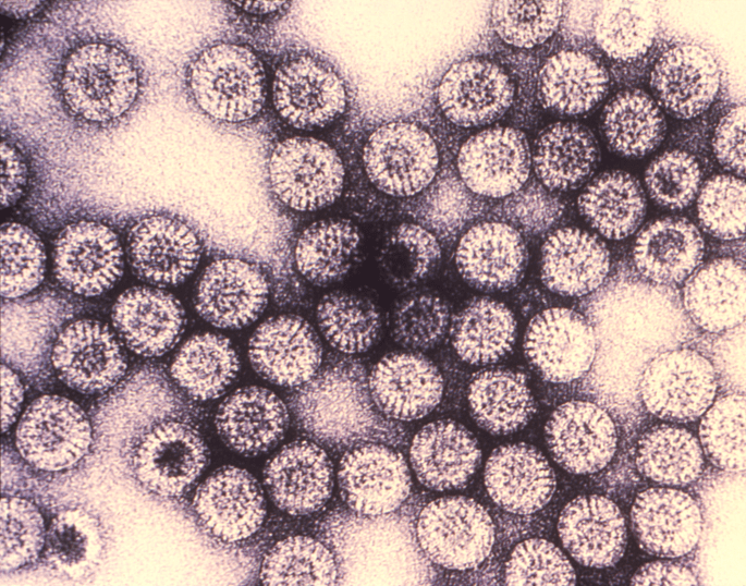
Under a very high magnification of 455,882X, this transmission electron micrograph (TEM) revealed some of the ultrastructural morphology exhibited by numbers of rotovirus icosahedral protein capsid particles.
Rotavirus disease is most common in infants and young children, but adults and older children can also become infected with rotavirus. Once a person has been exposed to rotavirus, it takes about 2 days for symptoms to appear.
Symptoms include:
– Fever
– Vomiting
– Diarrhea
– Abdominal pain
Vomiting and watery diarrhea may last from 3 to 8 days in a child who is infected with rotavirus. Additional symptoms include loss of appetite and dehydration (loss of body fluids), which can be especially harmful for infants and young children.
Vaccinated and unvaccinated children may develop rotavirus disease more than once because there are many different types of rotavirus and because neither vaccine nor natural infection provides full immunity (protection) from future infections. Usually a person’s first infection with rotavirus causes the most severe symptoms.
Image Credit and Caption: CDC
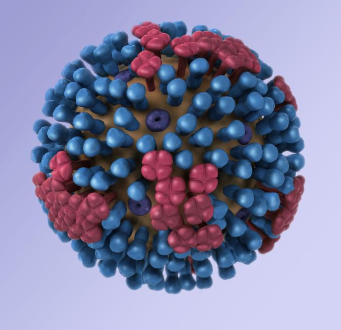
 This illustration provides a 3D graphical representation of a generic influenza virion’s ultrastructure, and is not specific to a seasonal, avian or 2009 H1N1 virus.
There are three types of influenza viruses: A, B and C. Human influenza A and B viruses cause seasonal epidemics of disease almost every winter in the United States. The emergence of a new and very different influenza virus to infect people can cause an influenza pandemic. Influenza type C infections cause a mild respiratory illness and are not thought to cause epidemics.
Influenza A viruses are divided into subtypes based on two proteins on the surface of the virus: the hemagglutinin (H), and the neuraminidase (N). There are 16 different hemagglutinin subtypes and 9 different neuraminidase subtypes. Influenza A viruses can be further broken down into different strains. Current subtypes of influenza A viruses found in people are influenza A (H1N1) and influenza A (H3N2) viruses. In the spring of 2009, a new influenza A (H1N1) virus emerged to cause illness in people. This virus was very different from regular human influenza A (H1N1) viruses and the new virus has caused an influenza pandemic.
Influenza B viruses are not divided into subtypes, however, influenza B viruses also can be further broken down into different strains.

From the CDC: “This photograph depicts the colonial growth pattern displayed by Salmonella typhimurium bacteria cultured on a Hektoen enteric (HE) agar medium; S. typhimurium colonies grown on HE agar are blue-green in color, for this organism is a lactose non-fermenter, but it does produce hydrogen sulfide, (H2S), therefore there can be black-colored deposits present.
HE agar is the medium designed for the isolation and recovery of fecal bacteria belonging to the family, Enterbacteriaceae. S. typhimurium causes 25% of the 1.4 million Salmonellosis infections a year in the United States. Most persons infected with Salmonella sp. develop diarrhea, fever, and abdominal cramps 12 – 72 hours after infection. The illness usually lasts 4 – 7 days, and most people recover without treatment. However, in some cases, the diarrhea may be so severe that the patient needs to be hospitalized.”
Image Credit: CDC

From the CDC: This scanning electron micrograph depicts a number of Gram-negative Campylobacter jejuni bacteria, magnified 9,951x.
Campylobacter is one of the most common bacterial causes of diarrheal illness in the United States. Virtually all cases occur as isolated, sporadic events, not as a part of large outbreaks with about 15 cases diagnosed each year for each 100,000 persons.
Image Credit: Janice Carr
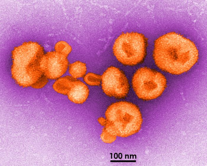
From the CDC: “This transmission electron micrograph depicted eight virions (viral particles) of a newly-discovered virus, which was determined to be a member of the genus, Arenavirus. A cause of fatal hemorrhagic fever, it was confirmed that this virus was responsible for causing illness in five South Africans, four of whom died having succumbed to its devastating effects.
Ultrastructurally, these round Arenavirus virions displayed the characteristic “sandy”, or granular capsid, i.e., outer skin, an appearance from which the Latin name, “arena”, was derived. See PHIL 10838 for a black and white version of this image.
Other members of the genus Arenavirus, include the West African Lassa fever virus, lymphocytic choriomeningitis (LCM), and Bolivian hemorrhagic fever (BHF), also known as Machupo virus, all of which are spread to humans through their inhalation of airborne particulates originating from rodent excrement, which can occur during the simple act of sweeping a floor.”
Image credit: CDC/Charles Humphrey
For those of you wondering what influenza’s mechanism of infection looks like, please see the below! Hemagglutinin is the H portion, and neuraminidase is the N – H1N1 therefore refers to the H1 hemagglutinin and the N1 neuraminidase. Both are surface glycoproteins responsible for host cell binding.
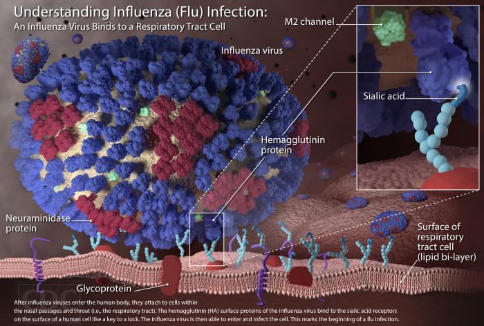
(Image: Arizona Department of Health)
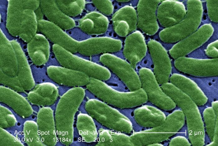
The image below is of Vibrio vulnificus, which as all of you undoubtedly know, is in the same family as Vibro cholerae.
From the CDC: ” [Vibrio vulnificus] normally lives in warm seawater and is part of a group of vibrios that are called ‘halophilic’ because they require salt. V. vulnificus can cause disease in those who eat contaminated seafood or have an open wound that is exposed to contaminated seawater. Among healthy people, ingestion of V. vulnificus can cause vomiting, diarrhea, and abdominal pain. In immunocompromised persons, particularly those with chronic liver disease, V. vulnificus can infect the bloodstream, causing a severe and life-threatening illness characterized by fever and chills, decreased blood pressure (septic shock), and blistering skin lesions. V. vulnificus bloodstream infections are fatal about 50% of the time.”
We shared this a couple years ago, but it’s making the internet rounds again, so we don’t feel bad re-posting it. Our image is:
Glass sculptures of pathogens!
Pictured below is H5N1, the strain of HPAI currently appearing in birds across China’s Guizhou province. The sculpture is done by artist Luc Jerram. Check out the rest of his gallery here.

This week’s image is this very cool infographic depicting the evolution of the zombie in film and video games.

