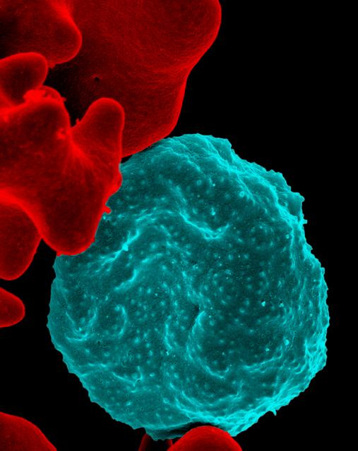
Pictured below is a red blood cell infected with malaria, false colored in blue – you can see the malaria virions as the brighter blue spots within the cells. To the left, uninfected red blood cells are shown in red, their surfaces smooth.
mala

Pictured below is a red blood cell infected with malaria, false colored in blue – you can see the malaria virions as the brighter blue spots within the cells. To the left, uninfected red blood cells are shown in red, their surfaces smooth.
mala
What does a chemical weapons inspector leaving to investigate chemical arsenals at a country in the middle of a raging civil war pack? Markers, apparently. The last thing they do before getting on said plane? You guessed it, fill out paperwork. Meet some of the men and women helping keep us out of Syria below.
(credit: OPCW)
Pictured below is H1N1, the pandemic virus better known (however unfairly) as “swine flu”. As many of you undoubtedly recall, H1N1 swept the globe in 2009, causing approximately 17,000 deaths.
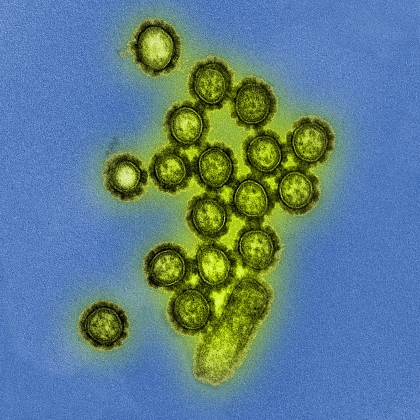
“Colorized transmission electron micrograph showing H1N1 influenza virus particles. Surface proteins on the virus particles are shown in black.” Image and caption: NIAID
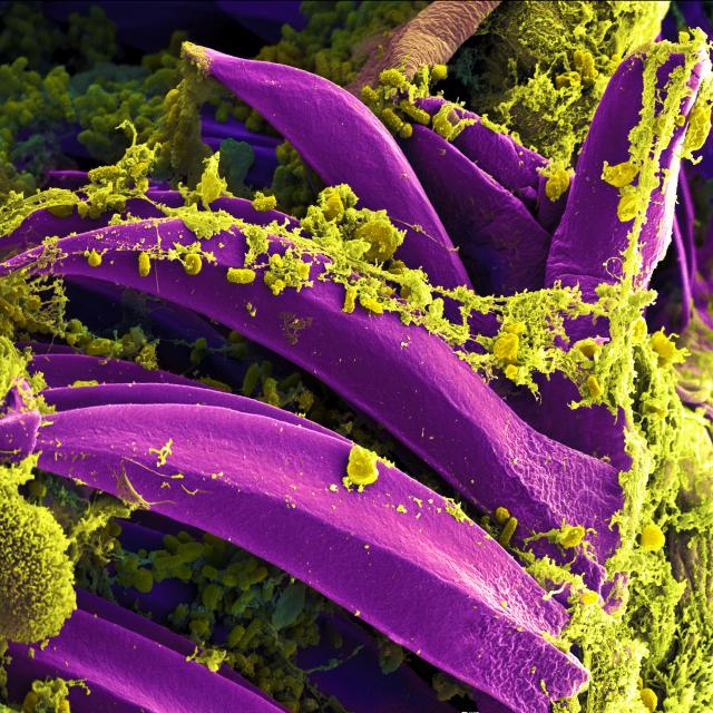
This week’s image shows us everyone’s favorite scourge – plague! Depicted below is Yersinia pestis, the causative agent of plague, sticking to the spines of a flea. Throughout history, Y. pestis has contributed to some of the most serious pandemics, at one point killing nearly half of Europe’s population. While plague still pops up occasionally (it’s endemic in the prairie dog population of the American Southwest), prompt treatment with antibiotics prevents serious illness. However, in aerosol form, unless antibiotics are administered in the first 24 hours, infection is almost always fatal. This is a big part of why Y.pestis is considered a potential bioterrorist agent.
Last week it was a chemical weapons slideshow (here, if you missed it), so this week we thought we’d swing back to something closer to our hearts – Ebola!
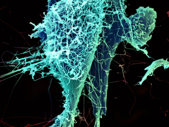
This rather startling image from NIAID shows Ebola particles “shedding” from infected cells, illustrating that Ebola is just as frightening on a molecular level. The virus’ distinctive, filamentous morphology is clearly visible.

Image of the Week: What does a chemical weapon look like? While we can’t show you the chemical weapons of Syria (we suggest youtube for that), we can show you images the CIA collected whilst doing analysis of the former Iraqi program. The images below focus on the weapons themselves, and are pulled almost exclusively from various CIA intelligence reports.
(header image: CIA)
This week’s image depicts the vacuole of a Vero cell, within which Coxiella burnetii, the bacteria which causes Q fever, is growing. Infection with just one of those green bacterium can cause Q fever in humans, an often asymptomatic disease which can occasionally result in respiratory symptoms, enlarged liver and spleen, and rarely, death. Q fever is found throughout the world, and is transmitted to humans primarily through interaction with contaminated livestock.
 (Image via NIAID/Flickr, click for larger image)

This beautiful image is of P. vortex, a bacteria species discovered in the ’90s by Eshel Ben-Jacob and his colleagues at the University of Tel Aviv. In an attempt to better understand the new species, Ben-Jacob tweaked various growth conditions, like temperature, light exposure, or nutrient consistency, and recorded the bacteria’s response. He soon found that the colony responded as a unit, resulting in distinctive patterns of movement and growth. Add a little dye, and boom – bacterial art.
Our favorite quote: “‘The bacteria have to maintain order, but they also have to maintain flexibility, so that when conditions change they can better adapt to the environment,’ says Ben-Jacob. ‘We have an affinity for things that have the combination of the two, order and disorder. If you analyze classical music, it is the same thing. The things that we really like and are captivated by are things that have this mixture.'”
To learn more and check out some of his other work, see here.
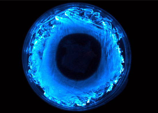
Three enterprising students from the University of Wisconsin, Madison are trying to create a light bulb powered entirely by E.coli. “But, wait!” You might say. “E.coli doesn’t bioluminesce!” In order to overcome this slight hurdle, the students plan on inserting a strip of DNA into the bacteria’s genome which will enable the bug to glow in the dark. The light bulb itself will contain the modified bacteria, the necessary growth medium, and a set of specific microbes capable of recycling light to provide nutrients and eliminate waste – all of which together would produce a bulb which glows without electricity, aptly dubbed Biobulb (pictured below).
Read more and fund the upstart project here!
(image courtesy of Cohn/Zaikan/Beckman, University of Wisconsin, Madison)
In the wake of the recent study detailing the first confirmed human to human transmission of H7N9 (Wired article here, for those of you who missed it), we thought we’d share this CDC infographic on the virus’ evolution.

